
Imaging
- Home
- our services
- Imaging

Multiple (Twins/Higher Order) Pregnancy Imaging
TIFFA Scan
Genetic Sonogram

Growth Scan
Fetal Echocardiography

Fetal Neurosonography

Fetal Doppler

Pelvis Scans

3D and 4D Imaging

Second Opinions
Nuchal Translucency (NT) & First Trimester Anatomy Scan
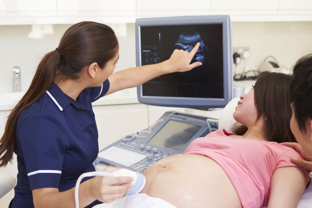
The Nuchal Translucency (NT) & First Trimester Anatomy Scan is a critical ultrasound performed between 11 and 14 weeks of pregnancy to evaluate early fetal development and screen for chromosomal abnormalities, such as Down syndrome (Trisomy 21). This non-invasive procedure measures the fluid accumulation at the back of the baby’s neck, examines the nasal bone, limbs, and major organs, and is often combined with maternal blood tests to estimate risk. Conducted by skilled sonographers, the scan typically takes 20–30 minutes and requires a moderately full bladder for optimal imaging. The results guide clinicians in planning timely follow-ups or additional testing, such as Non-Invasive Prenatal Testing (NIPT) or amniocentesis, ensuring early intervention if needed. This scan is a cornerstone of prenatal care, offering expectant parents peace of mind and valuable insights into their baby’s health.
Why Choose a Nuchal Translucency Scan?
Opting for an NT scan enhances early detection of potential developmental issues, allowing for proactive prenatal care. It’s a safe, routine procedure recommended for all pregnancies, particularly those with risk factors like advanced maternal age or a family history of chromosomal conditions. By identifying risks early, the NT scan supports informed decision-making and personalized care plans to ensure the best outcomes for both mother and baby.
Preparing for Your NT Scan
To prepare, drink water about an hour before the appointment to ensure a moderately full bladder, which improves ultrasound clarity. The procedure is painless and performed transabdominally, though a transvaginal approach may be used for better visualization in some cases. Discuss your medical history with your healthcare provider to maximize the scan’s effectiveness.
Multiple (Twins/Higher Order) Pregnancy Imaging
Multiple (Twins/Higher Order) Pregnancy Imaging is a specialized ultrasound designed to monitor the unique needs of twin or higher-order pregnancies. This advanced imaging tracks the growth and development of each fetus, evaluates the placenta, amniotic sacs, and umbilical cords, and detects complications like Twin-to-Twin Transfusion Syndrome (TTTS) or growth discrepancies. Regular scans assess cervical length, fetal well-being, and Doppler blood flow to ensure optimal health for both mother and babies. Frequent monitoring—often every 2–4 weeks—supports safer birth planning, timely interventions, and personalized care to minimize risks and improve outcomes.
Benefits of Multiple Pregnancy Imaging
Twins and higher-order pregnancies carry higher risks than singleton pregnancies, making specialized imaging essential. These scans provide detailed insights into each fetus’s health, helping detect issues early and guide decisions about delivery timing or specialized care. This proactive approach ensures safer pregnancies and healthier outcomes for both mother and babies.
What to Expect During the Scan
These ultrasounds are non-invasive and typically performed transabdominally. Depending on the pregnancy stage and complexity, scans may take 30–45 minutes. No special preparation is usually required, but your healthcare provider may recommend specific instructions based on your medical history.
TIFFA Scan
The Targeted Imaging for Fetal Anomalies (TIFFA) Scan, conducted between 18 and 22 weeks, is a comprehensive second-trimester ultrasound that examines the baby’s brain, heart, spine, face, abdomen, kidneys, limbs, and placenta. This high-resolution scan detects structural abnormalities, guides decisions about further genetic testing, and informs delivery planning. Recommended for all pregnancies, especially those with elevated risk from prior screenings, the TIFFA scan is safe, non-invasive, and critical for ensuring fetal health and preparing for any necessary interventions.
Importance of TIFFA Scans
The TIFFA scan plays a vital role in prenatal care by identifying anomalies early, allowing for timely management. It provides detailed images that help clinicians assess fetal development comprehensively, offering reassurance or guiding further diagnostics like amniocentesis. This scan is particularly valuable for high-risk pregnancies, ensuring tailored care for optimal outcomes.
How to Prepare for a TIFFA Scan
No special preparation is typically required, though a full bladder may enhance image clarity. The scan takes about 30–45 minutes and is performed by experienced sonographers using advanced ultrasound technology. Discuss any concerns or risk factors with your provider to ensure the scan addresses your specific needs.
Genetic Sonogram
A Genetic Sonogram is a specialized second-trimester ultrasound that uses “soft markers” and detailed anatomical checks to assess the risk of chromosomal abnormalities, such as Trisomy 21, 18, and 13. By examining features like the nuchal fold, nasal bone, heart anatomy, and long bones, this scan complements maternal blood tests and Non-Invasive Prenatal Testing (NIPT). The results help clinicians determine the need for further diagnostics, such as amniocentesis, and tailor prenatal care to ensure the best outcomes for mother and baby.
Why Get a Genetic Sonogram?
This scan enhances the accuracy of chromosomal risk assessments, providing critical information for high-risk pregnancies. It’s non-invasive, safe, and helps expectant parents make informed decisions about additional testing or specialized care. The genetic sonogram is particularly valuable when combined with other screening methods for a comprehensive evaluation.
What to Expect
The procedure is typically performed transabdominally and takes 20–30 minutes. No special preparation is needed, but discussing your family history or prior screening results with your provider can help customize the scan. Results are reviewed with your doctor to plan the next steps in your prenatal care journey.
Growth Scan
A Growth Scan, typically performed in the late second or third trimester, monitors fetal growth, amniotic fluid levels, placenta health, and baby’s position. Using biometric measurements like head circumference (HC), abdominal circumference (AC), femur length (FL), and estimated fetal weight (EFW), it identifies conditions like intrauterine growth restriction or macrosomia. Doppler studies may be included to assess blood flow and fetal well-being, guiding nutritional advice, medication adjustments, or early delivery planning for optimal outcomes.
Benefits of Growth Scans
Regular growth scans are essential for high-risk pregnancies, such as those with maternal diabetes, hypertension, or reduced fetal movements. They ensure timely detection of growth issues, enabling interventions to support healthy fetal development and safe delivery. These scans provide reassurance and critical data for personalized care plans.
Preparing for a Growth Scan
No specific preparation is required, though staying hydrated can aid image clarity. The scan takes about 20–30 minutes and is non-invasive. Your healthcare provider will review the results to discuss any necessary adjustments to your prenatal care or delivery plan.
Fetal Echocardiography
Fetal Echocardiography is a specialized ultrasound that examines the baby’s heart between 18 and 24 weeks, or whenever a cardiac concern arises. It assesses heart chambers, valves, outflow tracts, and rhythm to detect congenital heart defects early. Recommended for pregnancies with abnormal screenings, family history of heart disease, maternal diabetes, or certain medication exposures, this scan enables coordinated care with pediatric cardiologists and optimized delivery planning for better outcomes.
Why Fetal Echocardiography Matters
Early detection of heart defects allows for timely interventions, such as surgical planning or specialized monitoring post-birth. This non-invasive scan provides detailed images of the fetal heart, offering peace of mind or actionable insights for high-risk pregnancies.
What to Expect During the Procedure
The scan is performed transabdominally and may take 30–60 minutes, depending on complexity. No special preparation is needed, but discussing your medical history with your provider ensures a thorough evaluation. Results guide next steps, including referrals to specialists if needed.
Fetal Neurosonography
Fetal Neurosonography is an advanced ultrasound that provides a detailed examination of the fetal brain and spine. Recommended when TIFFA scans suggest neurological concerns, in high-risk pregnancies, or after infections like cytomegalovirus, this scan uses specialized views to detect neural tube defects or brain anomalies. Early identification supports further imaging, such as fetal MRI, and informs multidisciplinary care plans for optimal management and outcomes.
Importance of Fetal Neurosonography
This scan is critical for detecting complex neurological issues early, enabling timely counseling and planning. It’s particularly valuable for pregnancies with risk factors like maternal infections or genetic predispositions, ensuring comprehensive care tailored to the baby’s needs.
Preparing for the Scan
The procedure is non-invasive, typically transabdominal, and takes 30–45 minutes. No specific preparation is required, but sharing relevant medical history with your provider enhances the scan’s effectiveness. Results are discussed to determine next steps, including additional diagnostics if necessary.
Fetal Doppler
Fetal Doppler studies measure blood flow in critical vessels like the umbilical artery, middle cerebral artery (MCA), and uterine arteries to assess oxygen and nutrient delivery to the fetus. Essential for pregnancies with growth restriction, maternal hypertension, diabetes, or reduced fetal movements, Doppler scans guide surveillance frequency, medication adjustments, and delivery timing to protect fetal well-being and ensure healthy outcomes.
Benefits of Fetal Doppler Studies
Doppler ultrasounds provide real-time data on fetal circulation, enabling early detection of issues like placental insufficiency. This non-invasive test supports proactive interventions, reducing risks and optimizing maternal and fetal health, especially in high-risk pregnancies.
What to Expect
The procedure is quick, typically lasting 15–30 minutes, and is performed transabdominally. No preparation is needed, but staying hydrated can improve image quality. Your provider will review the results to adjust your care plan, ensuring the best possible outcomes.
Pelvis Scans
Pelvis Scans evaluate the uterus, cervix, ovaries, and adnexa to support pre-pregnancy planning, early pregnancy confirmation, and labor preparation. By measuring cervical length and assessing uterine anomalies or fibroids, these scans help predict risks like preterm birth or obstructed labor. The findings inform personalized antenatal care and delivery strategies, ensuring safer outcomes for mother and baby.
Why Pelvis Scans Are Important
These scans are crucial for identifying structural issues that could impact pregnancy or delivery. They’re recommended for women with a history of miscarriage, preterm birth, or pelvic abnormalities, providing critical data to guide clinical decisions and improve pregnancy outcomes.
Preparing for a Pelvis Scan
A full bladder may be required for optimal imaging, so drink water before the appointment. The scan is non-invasive, typically takes 15–30 minutes, and is performed transabdominally or transvaginally. Discuss your medical history with your provider to ensure a thorough evaluation.
3D and 4D Imaging
3D and 4D Imaging provides detailed, lifelike views of the baby’s face and anatomy, enhancing visualization of potential abnormalities and fostering parental bonding. While primarily complementary to diagnostic scans, these advanced ultrasounds clarify complex structures difficult to assess in 2D. Safe and non-invasive, they’re most effective between 24 and 32 weeks when amniotic fluid and fetal position optimize image quality, offering both medical insights and emotional connection.
Benefits of 3D and 4D Imaging
These scans offer clearer images than traditional 2D ultrasounds, aiding in the detection of subtle abnormalities and enhancing diagnostic accuracy. They also provide expectant parents with a unique bonding experience, making them a popular choice for both medical and emotional reasons.
What to Expect During the Scan
The procedure is non-invasive, typically takes 20–30 minutes, and requires no special preparation. Optimal timing depends on fetal position and amniotic fluid levels, so your provider may schedule the scan accordingly. Results are shared to enhance your prenatal experience and inform medical planning if needed.
Second Opinions
Second Opinions offer an expert review of prior ultrasound images and reports to confirm findings, clarify uncertainties, and guide next steps. Our specialists meticulously reassess image quality, measurements, and clinical context to ensure diagnostic accuracy and reduce parental anxiety. This service provides a clear, documented interpretation and a practical plan for follow-up testing, further imaging, or delivery planning, empowering expectant parents with confidence and clarity.
Why Seek a Second Opinion?
A second opinion can validate or refine initial findings, particularly in complex or high-risk pregnancies. It’s an invaluable tool for ensuring accuracy, addressing concerns, and making informed decisions about your prenatal care, especially when results are unclear or conflicting.
How the Process Works
Submit your prior ultrasound images and reports to our team for review. Our specialists will provide a detailed analysis, often within a few days, and discuss the findings with you. This service is designed to complement your existing care, offering peace of mind and actionable recommendations.